Dentine and Pulp 2
Dentine primary and secondary curvature of tubules, primary and secondary dentine, dead tract
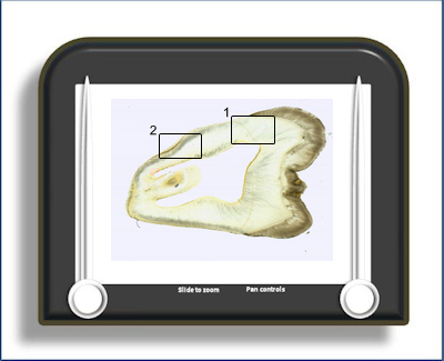
This is a ground longitudinal section of a tooth highlighting the coronal dentine (area 1) and root dentine (area 2). The dentine can be divided into either primary or (regular) secondary dentine. Or alternatively in the crown into mantle and circumpulpal dentine (the latter includes the primary dentine minus the mantle dentine plus the secondary dentine)
The mantle dentine is the first formed dentine which is laid down adjacent to the ADJ. The collagen fibres within it are predominantly arranged perpendicular to the ADJ (unlike the circumpulpal dentine) and are thought by some to arise not from odontoblasts but from early sub-odontoblastic mesenchyme. The evidence for this comes from the presence of von Korff fibres which are evident when the developing tooth is stained with silver.
The primary dentine is that dentine which is laid down to give the tooth its basic shape. Once the tooth has erupted and the root is completed there then follows a slower rate of dentine production at the pulpal surface throughout the life of the tooth, the regular secondary dentine (green arrow below). The junction between the two is usually marked by a change in direction of the dentinal tubules.
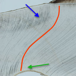
The dentinal tubules (particularly in the crown,
area 1)
do not
 pass
in a straight line from ADJ to pulpal surface
but follow a sinusoidal path called the primary
curvature (red line). At the highest magnification smaller
deviations in the path of the tubules can be
seen superimposed on the primary curvature -
these are the secondary curvatures. In the root
(area 2) the primary curvature is much less
evident.
pass
in a straight line from ADJ to pulpal surface
but follow a sinusoidal path called the primary
curvature (red line). At the highest magnification smaller
deviations in the path of the tubules can be
seen superimposed on the primary curvature -
these are the secondary curvatures. In the root
(area 2) the primary curvature is much less
evident.
In this ground section the path of some of the tubules is more obvious because they appear black. This is because they are empty (fluid filled and have no cell process or mineral deposit)and fill with debris during the grinding process - which produces the black appearance. In an area of dentine where a number of odontoblasts have been affected by caries, for instance, there may be a large and prominent group of tubules which appear black - this area is termed a 'dead tract' (blue arrow) (see area 1). The granular layer of Tomes and the 'hyaline' layer are present in area 2.
To open the e-Scope, click on one of the demarcated areas in the micrograph below:-