Dentine and Pulp
Dentine may be studied in both ground sections (where the mineral is retained and the section is unstained) and demineralised sections (where the mineral is removed and the section is stained) whereas pulp can only satisfactorily be studied in demineralised sections .
A characteristic feature of dentine is the presence of channels running through it from the pulp to the outer dentine surface – these are called dentinal tubules. These tubules contain fluid and a long process from the cells responsible for forming and maintaining the dentine – the odontoblasts. The tubules are not straight. They follow an overall ‘S’ shaped path called the ‘primary curvature’. Superimposed on this ‘long wavelength’ curvature are much more frequent, small wave-like deviations called the secondary curvature. The tubules may also be branched, particularly evident at the amelo-dentinal junction immediately under the enamel in the tooth crown.
 There
are three slides available to study the various
aspects of dentine and pulp structure and one
slide demonstrating the reactionary structures
in dentine resulting from an overlying enamel
caries lesion. Three
ground sections and one demineralised and
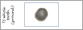
stained. Slide 1 is a ground section where the
tooth is sectioned in a transverse plane through
the enamel and dentine (this is the same section
available to view structures in enamel). On this
slide there are examples of interglobular
dentine, tubule branching and secondary
curvatures of the tubules. Slide 2 is also a
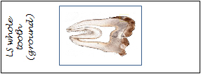
ground section but in the longitudinal plane
where primary and secondary tubule curvatures,
tubule branching, secondary dentine, dead
tracts, the granular layer of Tomes and the
hyaline layer are evident. Slide 3 is a demineralised
and stained transverse section through a root
showing dentine, the pulp/dentine interface and
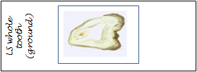
the pulp. Slide 4 is a longitudinal ground
section of a tooth showing sclerotic and
reparative dentine and dead tracts.
There
are three slides available to study the various
aspects of dentine and pulp structure and one
slide demonstrating the reactionary structures
in dentine resulting from an overlying enamel
caries lesion. Three
ground sections and one demineralised and
stained. Slide 1 is a ground section where the
tooth is sectioned in a transverse plane through
the enamel and dentine (this is the same section
available to view structures in enamel). On this
slide there are examples of interglobular
dentine, tubule branching and secondary
curvatures of the tubules. Slide 2 is also a
ground section but in the longitudinal plane
where primary and secondary tubule curvatures,
tubule branching, secondary dentine, dead
tracts, the granular layer of Tomes and the
hyaline layer are evident. Slide 3 is a demineralised
and stained transverse section through a root
showing dentine, the pulp/dentine interface and
the pulp. Slide 4 is a longitudinal ground
section of a tooth showing sclerotic and
reparative dentine and dead tracts.
Slide Box
2. A ground longitudinal section of a tooth demonstrating a number of aspects of dentine structure
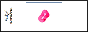
3. A demineralised transverse section of the dentine pulp interface