Periodontal ligament/Bone
The periodontium (the tooth support structure) has four constituent connective tissues, two of which are calcified and two which are non calcified. The two non-calcified (soft) connective tissues are the periodontal ligament (PDL), which fills the periodontal space between the root and the socket wall, and the lamina propria of the gingiva. By convention the boundary between the two is a horizontal line drawn at the level of the alveolar crest. The two calcified tissues are the cementum on the root surface (a bone-like material which can be either acellular or cellular) and the alveolar bone of the socket wall. This bone may also be referred to as the lamina dura (because it appears as a prominent white line on x-ray) or the cribriform plate (because it is perforated with numerous channels containing blood vessels). All four connective tissue elements of the periodontium share the same major matrix element: type I collagen.
There
are five slides available covering various
aspects of
 periodontal and alveolar bone
structure. Slide 1 is a demineralised section of
a tooth in situ showing periodontal structure,
bone formation (osteoid) and bone resorption.
Slide 2 is also demineralised but is stained
with picric acid (rather than H&E) - this
highlights the collagen fibres, especially as
they insert into bone and cementum. Slide 3 is
demineralised but then pre-oxidised before
staining with an elastin stain - this stains
the oxytalan fibres within the PDL. Slide 4 is a
(micro)radiograph of a ground section of a tooth
in its socket - this demonstrates the varying
degrees of mineralisation within the bone of the
socket of the tooth. Slide 5 is a ground section
of a tooth highlighting the structural features
of cementum.
periodontal and alveolar bone
structure. Slide 1 is a demineralised section of
a tooth in situ showing periodontal structure,
bone formation (osteoid) and bone resorption.
Slide 2 is also demineralised but is stained
with picric acid (rather than H&E) - this
highlights the collagen fibres, especially as
they insert into bone and cementum. Slide 3 is
demineralised but then pre-oxidised before
staining with an elastin stain - this stains
the oxytalan fibres within the PDL. Slide 4 is a
(micro)radiograph of a ground section of a tooth
in its socket - this demonstrates the varying
degrees of mineralisation within the bone of the
socket of the tooth. Slide 5 is a ground section
of a tooth highlighting the structural features
of cementum.
Slide Box
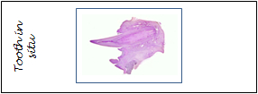
1. Tooth in situ - PDL and bone (H&E)
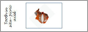
2. Tooth in situ - PDL and bone (picric acid)
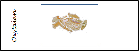
3. Tooth in situ - Oxytalan fibres in PDL (pre-oxidised slide)
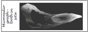
4. Tooth in situ - microradiograph
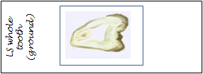
5. Ground section of tooth showing cementum