Oral Mucosa
Areas of the oral mucosa can be classified according to their structure or their function, although the two are intimately related. All areas of the mucosa share the same basic underlying structure but which is more or less modified to perform the required function. The outer layer is the epithelium (which may or may not be keratinised), then a dense connective tissue layer - the lamina propria. Beneath this there may be a looser region of connective tissue and vascular tissue - the sub mucosa. Finally the mucosa will ultimately sit upon a bed of either bone or muscle.
Lining mucosae (usually) have a relative thick non-keratinised epithelium with broad, shallow epithelial ridges/dermal papillae. A loose sub-mucosa is present.
Masticatory mucosae have keratinised epithelia which may be ortho or para keratinised (where some cell nuclei may be still present at the surface), the keratin presenting as a more intensely stained outer layer (sometimes with a more orange hue). The epithelial ridges/dermal papillae are long and pointed. There is no submucosa, the lamina propria attaching to the underlying bone - termed a mucoperiosteum.
Specialised mucosae have extreme modifications of these basic forms to perform specific functions.
The lining mucosae are:- inner surface of the lip, the alveolar mucosa, the oral surface of the soft palate, the underside of the tongue/floor of the mouth and the cheek.
The masticatory mucosae are:- the gingiva and the hard palate .
The specialised mucosae are :- the dorsum of the tongue, the vermillion border of the lip and the outer (hairy) skin of the lip.
 There are seven slides available to cover the
regions of the oral mucosa. All slides are of
demineralised tissue, stained with H&E. The
seven slides are:
Slide Box
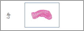
1. Lip (inner and outer surface and vermillion border)
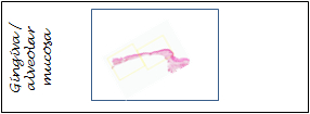
2. Gingiva and alveolar mucosa
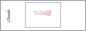
3. Cheek
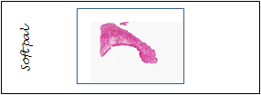
4. Soft palate (oral and nasal surfaces)
5. Hard palate
