Enamel
Enamel is a very brittle and hard-wearing material and to a very great extent relies on the underlying dentine for resilience.
It is almost pure mineral – only approximately 4% is water and organic material which is mostly (non-collagenous) protein. The mineral is hydroxyapatite (the same as that in bone and dentine, although the apatite crystals in enamel are much larger). The enamel forms a hard-wearing covering over the dentine of varying thickness, varying from its thickest extent at the incisal edge(or cusp in a molar tooth) to a knife-edge thinness at the cervical margin.
The crystals of hydroxyapatite are not randomly arranged but ordered into assemblies called prisms (also called rods). Each prism runs from the dentine to just below the tooth surface and is approximately 5µm in diameter. In longitudinal sections of the tooth the prisms are approximately perpendicular to the amelo-dentinal junction. There is a circadian rhythm in enamel production which produces regular, daily, cross-striations with roughly a 5µm periodicity. Superimposed on this periodicity are other less frequent incremental lines called the (brown) striae of Retzius. Towards the cusp these tend to be irregularly spaced but towards the cervical margin they are much more regular.
There are other structural features in enamel not directly related to prism structure. Some arise from events in the formation of the enamel and others are essentially optical effects produced by the overall arrangement of the prisms. Some of these are associated with the amelo-dentinal junction and others refer to areas or features within the bulk of the enamel.
 There are two slides available to look at
structures in enamel and one demonstrating a
'white spot' caries lesion. All three slides are ground
sections of whole teeth. It is necessary to use
ground sections because, as enamel is
essentially all mineral, demineralisation would
completely remove it, leaving an enamel 'space'.
Slide 1 is a transverse section of a tooth
showing enamel tufts emanating from the amelo-dentinal
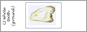
junction. Slide 2 is a longitudinal section
where a number of other enamel structures can be
studied. Slide 3 is a longitudinal section with
a smooth surface white spot lesion.
There are two slides available to look at
structures in enamel and one demonstrating a
'white spot' caries lesion. All three slides are ground
sections of whole teeth. It is necessary to use
ground sections because, as enamel is
essentially all mineral, demineralisation would
completely remove it, leaving an enamel 'space'.
Slide 1 is a transverse section of a tooth
showing enamel tufts emanating from the amelo-dentinal
junction. Slide 2 is a longitudinal section
where a number of other enamel structures can be
studied. Slide 3 is a longitudinal section with
a smooth surface white spot lesion.
Slide Box
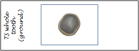
1. A ground transverse section of a tooth showing enamel tufts
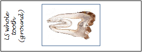
3. A ground longitudinal section of a tooth showing a smooth surface white spot lesion