Enamel 3
White spot lesion
This is a ground longitudinal section through a whole tooth which has several carious lesions in both the enamel and dentine. This particular tooth demonstrates the classical conical shape of a smooth surface, white spot, lesion (the region demarcated) and also a fissure lesion where the visible lesion is much smaller than the lesion in the underlying enamel and dentine. This is largely a result of the divergent path of the prisms away from the fissure. The smooth surface lesion demonstrates the classic zones - translucent zone, dark zone surface zone and lesion body. Because of the partial demineralisation of the enamel, the structural elements within it (i.e. the striae of Retzius and the cross-striations) appear more distinct than in normal enamel.
The translucent zone: This zone appears translucent because it takes up imbibing media into relatively uniformly sized pores produced by the earliest stages of acid dissolution. It has lost approximately 1% of its mineral.
The body of the lesion: The lesion body is said to have lost >20% of its mineral.
The dark zone: Some remineralisation has occured within this zone so that the pores (produced by the initial demineralisation) demonstrate a range of sizes, some of which are relatively small and inhibit the ingress of the imbibing fluid. This causes scattering of the light and hence the dark colouration. This zone may also contain a relatively high protein content. This zone is said to have lost approximately 5% of its mineral.
The surface zone: A relatively intact surface zone is characteristic of this lesion type so that demineralisation is apparently sub-surface. The presence of this relatively intact zone appears to be independent of fluoride concentration. Micro channels may be present passing through this zone when viewed at sufficiently high power.
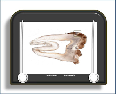
To open the e-Scope, click on the demarcated area in the micrograph below:-