Enamel 1
Tufts and spindles
If the enamel of the crown is completely removed with acid (demineralised), ribbons of protein can often be seen to run down the exposed dentine surface – this is tuft protein and represents areas between prisms where residual enamel protein has collected.
The section presented here is a transverse
ground
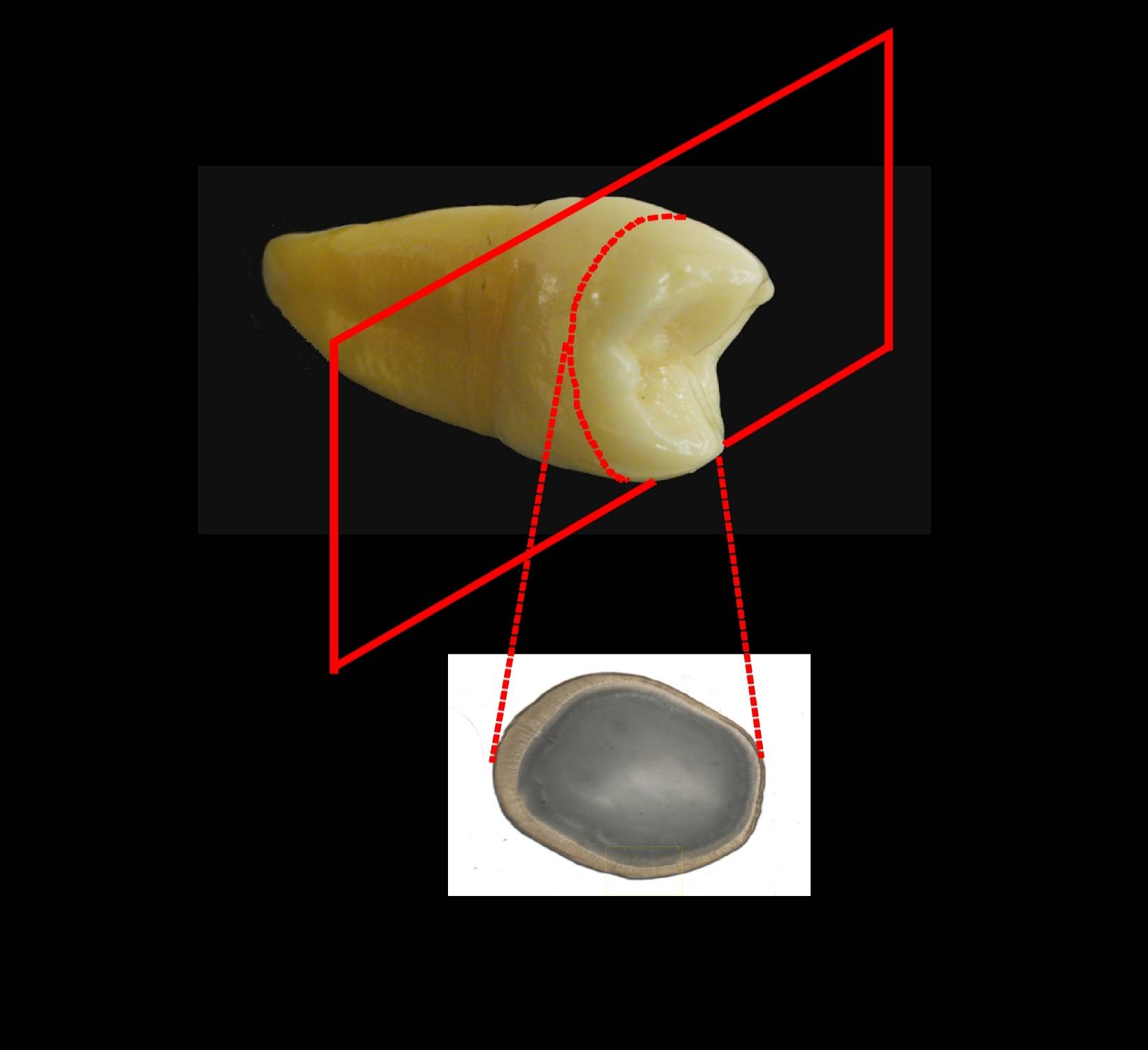 (undemineralised) thick section of a
tooth showing enamel and dentine either side of
the amelo-dentinal junction (ADJ) (which is
scalloped in appearance). The
structures visible in the enamel originating
from the ADJ are enamel tufts and enamel
spindles
(undemineralised) thick section of a
tooth showing enamel and dentine either side of
the amelo-dentinal junction (ADJ) (which is
scalloped in appearance). The
structures visible in the enamel originating
from the ADJ are enamel tufts and enamel
spindles
The prisms in the enamel do not run in a straight line in these transverse sections but take a sinusoidal path to the surface (unlike the straight line in longitudinal sections) with all the prisms at a given level running parallel to each other.
However,
a thick section such as this one and the one via
 the e-Scope contains
several prism levels within its thickness which
are superimposed. They are visible because of
the high protein content retained at the prism
boundaries and, because of the superimposition,
give the appearance of a tuft of grass (red
arrow) - hence the name.
the e-Scope contains
several prism levels within its thickness which
are superimposed. They are visible because of
the high protein content retained at the prism
boundaries and, because of the superimposition,
give the appearance of a tuft of grass (red
arrow) - hence the name.
Spindles
also originate at the ADJ and project into the
enamel.
 However, unlike the tufts, these are
discrete 'cigar' shaped structures and are only
clearly visible at one particular level within
the thick section (blue arrow).
To see a through focus series of an enamel tuft
- click here. Note that a
spindle is also visible but only at one level
within the thick section and will go in and out
of focus. Individual tufts will be visible
throughout the section thickness but will change
their appearance as the different prism levels
are brought into focus.
However, unlike the tufts, these are
discrete 'cigar' shaped structures and are only
clearly visible at one particular level within
the thick section (blue arrow).
To see a through focus series of an enamel tuft
- click here. Note that a
spindle is also visible but only at one level
within the thick section and will go in and out
of focus. Individual tufts will be visible
throughout the section thickness but will change
their appearance as the different prism levels
are brought into focus.
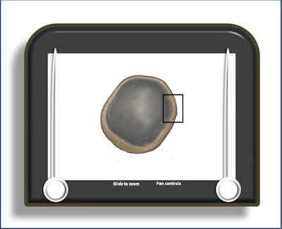
To open the e-Scope, click on the demarcated area in the micrograph below:-