Tooth Development 1
Bell stage (+ bud of permanent tooth)
The change from the 'cap' stage to the 'bell' stage is a gradual one and not a sudden jump from one to the other. The term 'bell' is derived from the shape of the basal lamina between the inner enamel epithelium and the outer cells of the papilla.
This is a saggital section through a foetal head showing both upper and lower incisors at the bell stage of development. The upper incisor is at a slightly later stage, however, being at the 'late' bell stage. The lower incisor is still in the 'early' bell stage. The significant difference between the two is that in the former the dentine matrix has begun to form at the cusp tip. In the lower incisor there is no evidence of matrix. Considerable morphodifferentiation (development of crown shape) and almost complete histodifferentiation have occurred.
A number of distinct tissue types are now evident. Those of the enamel organ will form the enamel of the tooth, the papilla will form the dentine and pulp and the follicle will form the periodontal ligament and cementum. The enamel organ is made up of a number of discrete cell populations (going from outer to inner):
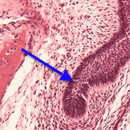
1. The
outer enamel epithelium (OEE) (blue
arrow) is a single
layer of
 small cuboidal cells which form the
outer envelope of the enamel organ and is
continuous with the inner enamel epithelium (see
below) at
the cervical loop. The OEE has lost its
continuity with the dental lamina by this stage.
At the onset of the late bell
stage, the stellate reticulum above the cusp
collapses so that the OEE is brought close the
the tissues at the cusp. The OEE also becomes
invaginated with capillaries at this point and
is often referred to as the 'papillary' layer.
small cuboidal cells which form the
outer envelope of the enamel organ and is
continuous with the inner enamel epithelium (see
below) at
the cervical loop. The OEE has lost its
continuity with the dental lamina by this stage.
At the onset of the late bell
stage, the stellate reticulum above the cusp
collapses so that the OEE is brought close the
the tissues at the cusp. The OEE also becomes
invaginated with capillaries at this point and
is often referred to as the 'papillary' layer.
2. The
stellate reticulum (SR) is an open
meshwork of star-shaped cells which form the
bulk of the enamel
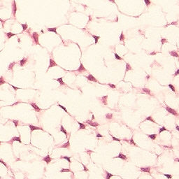 organ. These cells produce
large quantities of glycosaminoglycans which
bind water and help to make the enamel organ
turgid. This provides protection for the
developing tooth-forming cells and provides the
environment in which the correct cuspal pattern
can develop.
organ. These cells produce
large quantities of glycosaminoglycans which
bind water and help to make the enamel organ
turgid. This provides protection for the
developing tooth-forming cells and provides the
environment in which the correct cuspal pattern
can develop.
3. The
stratum intermedium is a layer of
flattened cells (blue
arrow), one or two cells thick, which
sits at the base of the internal enamel
epithelium cells (red arrow). It appears during the
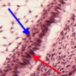 transition from cap to bell stage. It is rich in
alkaline phosphatase and appears necessary for
the terminal differentiation of ameloblasts from
the inner enamel epithelium and the production
of enamel - its absence in the root sheath
appears to result in the lack of enamel
production on the root.
transition from cap to bell stage. It is rich in
alkaline phosphatase and appears necessary for
the terminal differentiation of ameloblasts from
the inner enamel epithelium and the production
of enamel - its absence in the root sheath
appears to result in the lack of enamel
production on the root.
4. The histodifferentiation within the enamel organ has produced an 'inner lining' adjacent to the papilla termed the inner enamel epithelium (IEE) (red arrow). It consists of a single layer of cells which are noticeably more columnar than the outer enamel epithelium. At the bell stage there is clear evidence of differential development along the length of the IEE, with the cells being more developed (i.e. more columnar) at the developing cusp compared to the cervical loop where the IEE is continuous with the OEE.
The papilla is the (ecto)mesenchymal condensation (originally derived from the neural crest) which is enclosed by the enamel organ. It will eventually give rise to the odontoblasts (and hence the dentine) and the pulp of the tooth. At the early bell stage the cells of the papilla at the developing cusp (the odontoblasts) have elongated but have not yet produced dentine matrix. At the late bell stage the cells at the cusp tip have fully differentiated and have started to produce dentine matrix, the outer layer of which has begun to mineralise (as evidenced by the darker staining). Teeth at this stage would become visible on a clinical x-ray.
The dental follicle is the (ecto)mesenchyme which surrounds the enamel organ and papilla and will eventually give rise to the cementum, periodontal ligament and the bone of the socket wall. At bell stage, the follicle can clearly be divided into a denser region adjacent to the enamel organ, called the investing layer, and a looser more vascular region adjacent to the bone.
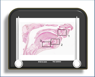
In the slide below area 1 is the upper incisor
at late bell stage
 and
area 2 is the lower incisor at early
bell stage. The bud of its permanent successor
(blue arrow)
is adjacent (lingual) to it. There has been a
significant amount of development of the
mandible by this stage and Meckel's
cartilage is reduced in size and is displaced by
the body of the mandible (area 3).
and
area 2 is the lower incisor at early
bell stage. The bud of its permanent successor
(blue arrow)
is adjacent (lingual) to it. There has been a
significant amount of development of the
mandible by this stage and Meckel's
cartilage is reduced in size and is displaced by
the body of the mandible (area 3).
To open the e-Scope, click on one of the demarcated areas in the micrograph below:-