Tooth Development 2
Cap stage
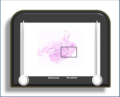
This is a saggital section through a foetal head showing a tooth at the 'cap' stage of development. There has been some development of the mandible by this stage with both lateral and medial plates of bone present and Meckel's cartilage is starting to reduce in size and is displaced by the growing body of the mandible. Some limited morphodifferentiation (development of crown shape) and histodifferentiation are evident.
The enamel organ (which forms the 'cap' shape) is the ectodermally derived portion of the tooth germ and, as its name suggests, will give rise to the cells that will produce the enamel. It consists, at this stage, of the outer enamel epithelium (OEE) and inner enamel epithelium (IEE) and a more loosely packed central core (which will eventually form the stellate reticulum -see bell stage slides).
The outer enamel epithelium consists of small cuboidal cells and is continuous with cells on the inner surface of the 'cap', the inner enamel epithelium, at a structure termed the cervical loop. The OEE is also still continuous with the dental lamina at this stage.
The histodifferentiation within the enamel organ has produced an 'inner lining' adjacent to the papilla termed the inner enamel epithelium (the papilla will eventually form the dentine and pulp of the tooth). It consists of a single layer of cells which are noticeably more columnar than the outer enamel epithelium. At the cap stage there is little evidence of differential development along the length of the IEE.
To open the e-Scope, click on the demarcated area in the micrograph below:-
