Periodontal ligament/Bone 5
Acellular and cellular cementum
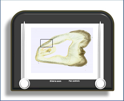
This slide is a ground section showing acellular and cellular cementum with some evidence of incremental lines (of Salter)
As the name implies, acellular cementum has no
cellular element. It is generally thinner than
cellular cementum
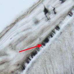 but has a wider distribution,
covering the whole of the root. It is the first
formed cementum. Characteristically in ground
sections it demonstrates parallel black lines,
perpendicular to the tooth surface (red arrow). These lines
result from the points of insertion of the
'extrinsic' fibres from the PDL into the cementum. The relatively low mineralisation at
the junction between 'intrinsic' and 'extrinsic'
fibres results in this black appearance due to
preferential grinding and debris collection at
these points.
but has a wider distribution,
covering the whole of the root. It is the first
formed cementum. Characteristically in ground
sections it demonstrates parallel black lines,
perpendicular to the tooth surface (red arrow). These lines
result from the points of insertion of the
'extrinsic' fibres from the PDL into the cementum. The relatively low mineralisation at
the junction between 'intrinsic' and 'extrinsic'
fibres results in this black appearance due to
preferential grinding and debris collection at
these points.
The bulk of the cementum, particularly at the root apex contains cells (cementocytes) hence the name 'cellular' cementum. It is usually deposited onto a layer of 'acellular' cementum and is deposited throughout the life of the tooth to accommodate for occlusal wear of the crown. It is a bone-like material although unlike bone it does not posses its own blood supply (obtaining its supply from the adjacent periodontal ligament and hence the polarisation of cementocyte canaliculi). Neither does it have bone's organisation into osteons.
The cementocyte is the cellular element of 'cellular' cementum. It is derived from a cementoblast which becomes entombed in cementum matrix. It is analogous to the osteocyte in bone. However, it differs in that its canaliculi extending from its containing lacuna are generally polarised towards the periodontal space. In bone they are arrayed more symmetrically. It has a reduced organelle content compared to the cementoblast.
Cementum demonstrates relatively irregular incremental growth lines parallel to the tooth surface - these are the incremental lines of Salter.
To open the e-Scope, click on the demarcated area in the micrograph below:-