Dentine and Pulp 3
Dentine/pulp interface
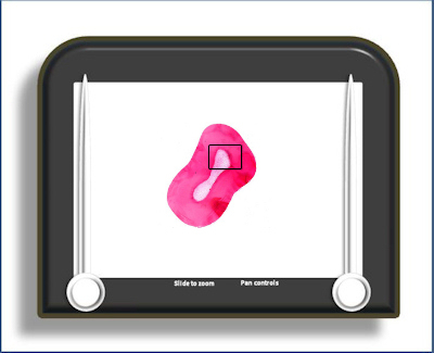
This is a demineralised transverse section through the root of a tooth showing pulp, dentine and the pulp/dentine interface.
The pulp dentine interface is traditionally described as a series of layers, all of which are visible on this slide. These are (from exterior to interior):
- mineralised dentine
- pre-dentine (unmineralised except for the presence of calcospherites)
- odontoblasts
- cell free zone (of Weil)
- cell rich zone
- body of the pulp (a loose, vascular and well innervated connective tissue)
Predentine (green
arrow) is the initial
collagenous matrix of dentine before it is
mineralised. There is always a
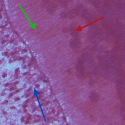 layer of predentine between the
odontoblast layer (blue arrow) and the mineralised
dentine. It is homologous to osteoid on the
surface of forming bone. The odontoblast is the cell
responsible for forming dentine and maintaining
it. It initially secretes a collagenous matrix (the predentine) which is
then subsequently mineralised (partly by the
fusion of calcospherites). It is a highly
polarised cell with prominent organelles for the
production and secretion of protein and a
basally placed nucleus. A process from the
distal end of the cell projects into a dentinal
tubule. As dentine production progresses and the
pulp becomes reduced in size the odontoblast
layer becomes pseudostratified. The organelle
count also decreases as the cell moves from the
rapid production of primary dentine to the
slower, long term, production of regular
secondary dentine. There are numerous cell
contacts between adjacent odontoblasts. The
odontoblast layer also contains unmyelinated
nerve fibres and numerous capillary loops, both
of which arise from a sub-odontoblastic plexus.
layer of predentine between the
odontoblast layer (blue arrow) and the mineralised
dentine. It is homologous to osteoid on the
surface of forming bone. The odontoblast is the cell
responsible for forming dentine and maintaining
it. It initially secretes a collagenous matrix (the predentine) which is
then subsequently mineralised (partly by the
fusion of calcospherites). It is a highly
polarised cell with prominent organelles for the
production and secretion of protein and a
basally placed nucleus. A process from the
distal end of the cell projects into a dentinal
tubule. As dentine production progresses and the
pulp becomes reduced in size the odontoblast
layer becomes pseudostratified. The organelle
count also decreases as the cell moves from the
rapid production of primary dentine to the
slower, long term, production of regular
secondary dentine. There are numerous cell
contacts between adjacent odontoblasts. The
odontoblast layer also contains unmyelinated
nerve fibres and numerous capillary loops, both
of which arise from a sub-odontoblastic plexus.
Calcospherites are small isolated globular areas of mineralisation within predentine which eventually fuse with the mineralisation front (and each other) (red arrow).
The cell free zone lies beneath the odontoblast layer and appears devoid of cells on this typical H&E stained section. However, at higher magnifications it can be seen that while it may not contain many cell nuclei it has numerous cell processes running through it. Also many small capillaries loops run through it as they pass from the sub-odontoblastic capillary plexus into the odontoblast layer. It is less obvious in radicular pulp. Many now consider the appearance of the cell-free zone to be artefactual, arising from shrinkage of the pulp tissue away from the odontoblast layer (which is attached firmly to the dentine) during processing.
As the name suggests, the cell rich zone (unlike the cell free zone) appears to contains a high concentration of cell nuclei.
The body of the pulp constitutes the loose connective tissue which occupies the space in the remainder of the pulp chamber. The predominant cell type is the fibroblast although macrophages may be present (more substantiatial changes in cell content will occur in the inflammed pulp). Undifferentiated mesenchyme is also present in a sub-odontoblastic location to provide cells to replace odontoblasts which may be damaged. The pulp also contains the nerves and capillaries which supply the odontoblast layer (as well as the pulp itself) - these enter the pulp through the apical foramen. Many of the nerves will have endings between odontoblasts or within dentinal tubules. The capillaries form numerous loops within the odontoblast layer and may be fenestrated.
To open the e-Scope, click on the demarcated area in the micrograph below:-