Oral Mucosa 6
Dorsum of tongue
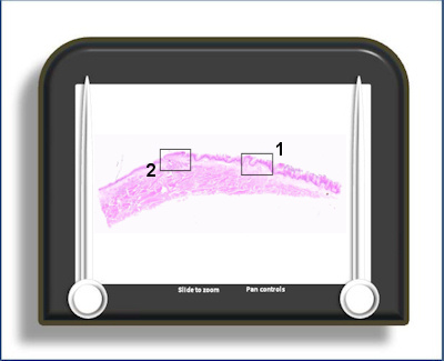
This slide is of the upper surface of the tongue showing the structures which give the surface its roughness (filiform papillae) and also those which contain taste buds (fungiform and circumvallate papillae).
Area 1 shows filiform
papillae (blue arrow)
and a single
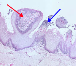 fungiform
papilla (red arrow).
The filiform papillae provide the surface
roughness of the tongue because the keratin
layer projects vertically from the surface. In a
normal keratinised epithelium the surface
keratinised cells lie parallel to the surface
which is essentially smooth as a result. The
cells are produced in the basal layer on the
lateral borders of the papilla. They then
migrate outwards then upwards orientated at
roughly 90o to the surface. The fungiform
papillae are much less numerous than the
filiform papillae. They have a mainly
non-keratinised epithelium with taste buds on
their lateral borders (which are much less
frequent than on the circumvallate papillae). As
this section shows, they have a well developed
blood supply which comes close to the
epithelium. This proximity to the surface gives
these papillae a red colouration compared to the
paler filiform papillae.
fungiform
papilla (red arrow).
The filiform papillae provide the surface
roughness of the tongue because the keratin
layer projects vertically from the surface. In a
normal keratinised epithelium the surface
keratinised cells lie parallel to the surface
which is essentially smooth as a result. The
cells are produced in the basal layer on the
lateral borders of the papilla. They then
migrate outwards then upwards orientated at
roughly 90o to the surface. The fungiform
papillae are much less numerous than the
filiform papillae. They have a mainly
non-keratinised epithelium with taste buds on
their lateral borders (which are much less
frequent than on the circumvallate papillae). As
this section shows, they have a well developed
blood supply which comes close to the
epithelium. This proximity to the surface gives
these papillae a red colouration compared to the
paler filiform papillae.
Area 2 contains a
circumvallate papilla (red
arrow) with a minor
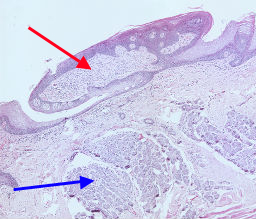 (serous)
salivary gland (of von Ebner) beneath it (blue
arrow) and taste buds on its side wall.
The circumvallate papillae are arranged in the
form of a 'V' at the junction between the
anterior 2/3rds and posterior 1/3rd of the
tongue at the sulcus terminalis. They have
numerous taste buds on their lateral borders.
They sit below the tongue surface in an
encircling depression. This is constantly
irrigated by a serous secretion of the von Ebner
glands. These are the only 'serous' minor
salivary glands. Without this constant flushing
of the area food substances would remain,
blocking the possibility of tasting new taste
stimuli during eating.
(serous)
salivary gland (of von Ebner) beneath it (blue
arrow) and taste buds on its side wall.
The circumvallate papillae are arranged in the
form of a 'V' at the junction between the
anterior 2/3rds and posterior 1/3rd of the
tongue at the sulcus terminalis. They have
numerous taste buds on their lateral borders.
They sit below the tongue surface in an
encircling depression. This is constantly
irrigated by a serous secretion of the von Ebner
glands. These are the only 'serous' minor
salivary glands. Without this constant flushing
of the area food substances would remain,
blocking the possibility of tasting new taste
stimuli during eating.
To open the e-Scope, click on one of the the demarcated areas in the micrograph below:-