Enamel 2
Striae of Retzius, gnarled enamel, spindles, cross-striations, Hunter Schreger bands
This is a longitudinal ground section of a
tooth. The
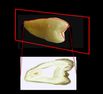 section is unstained but in
transmitted light the enamel has taken on a
brown hue. Several structural details of enamel
are evident in this section - notably the Striae
of Retzius, enamel spindles (also seen in enamel
in TS), the 'scalloped' nature of the amelo-dentinal
junction (ADJ) itself, the so-called 'gnarled'
enamel, Hunter-Schreger bands and
cross-striations of the enamel prisms.
section is unstained but in
transmitted light the enamel has taken on a
brown hue. Several structural details of enamel
are evident in this section - notably the Striae
of Retzius, enamel spindles (also seen in enamel
in TS), the 'scalloped' nature of the amelo-dentinal
junction (ADJ) itself, the so-called 'gnarled'
enamel, Hunter-Schreger bands and
cross-striations of the enamel prisms.
The (brown) striae of Retzius are incremental
growth lines that on the sides of the crown run
obliquely across
 the prisms to approach the
enamel surface. At the surface they are often
associated with a depressions called perikymata.
Particularly in the outer regions of the enamel
and towards the cervical margin of the crown (as
in this case) they may be very regularly spaced,
approximating to seven days growth (area 2). At the cusps
the striae run around the cusp and do not
approach the surface and appear similar to tree
rings (area 1). Striae mark the forming surface of the
enamel at a particular time point in the crown's
development.
the prisms to approach the
enamel surface. At the surface they are often
associated with a depressions called perikymata.
Particularly in the outer regions of the enamel
and towards the cervical margin of the crown (as
in this case) they may be very regularly spaced,
approximating to seven days growth (area 2). At the cusps
the striae run around the cusp and do not
approach the surface and appear similar to tree
rings (area 1). Striae mark the forming surface of the
enamel at a particular time point in the crown's
development.
The ADJ is the junction between the enamel and dentine. It has a characteristic 'scalloped' appearance. This scalloping aides the retention of the enamel on the dentine and resists shearing forces by providing a more irregular and greater surface area at the point of contact.
The enamel spindle is a cigar-shaped structure which can often be seen to be continuous with dentinal tubules across the ADJ. They are thought to arise because of growth of odontoblast processes across the forming ADJ into the developing enamel during crown development (can be seen in both areas 1 and 2)
On the side of the crown the enamel prisms run in essentially straight lines to the surface at an angle approximating 90o to the ADJ (area 2). However, at the cusp tip (mid region of area 1) the prisms run in what appears to be a much more disordered fashion - this is the so-called 'gnarled' enamel and has evolved to resist the greater loads and therefore shearing forces at the cusp. (It has been suggested that the apparent 'disorder' is, in fact, an artefact of sectioning through a more complicated [but ordered] arrangement at the cusp - possibly a spiral).
Particularly in area 1 of this section, at the highest magnification, the cross-striation in the enamel prisms can be seen - these represent daily incremental growth lines and give the prism a ladder-like appearance.
Also visible in area 1 are examples of Hunter-Schreger bands where groups of prisms run at slightly different paths to the surface (this arrangement gives rise to the tufts seen in transverse sections of enamel).
To open the e-Scope, click on one of the demarcated areas in the micrograph below:-
