Oral Mucosa 1
Lip
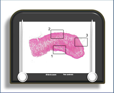
This is a longitudinal section of the lip, stained with H&E. It has three areas of interest which can be viewed in the microscope: the inner, lining mucosa (towards the bottom of the slide) (area 1), the outer, hairy, skin towards the top of the slide (area 2) and the vermillion border (the red area of the lip) towards the right hand side of the slide (area 3).
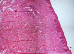
Area
1 is a view of the mucosa on the inner surface of the lip. It
 is a typical lining mucosa with a non keratinised epithelium, short and rounded epithelial ridges and a sub mucosa.
A minor salivary gland is situated in the submucosa and underlying muscle.
is a typical lining mucosa with a non keratinised epithelium, short and rounded epithelial ridges and a sub mucosa.
A minor salivary gland is situated in the submucosa and underlying muscle.
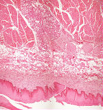
Area
2 shows the surface layers of the outer
surface of the lip
 (hairy skin). It is a
keratinised epithelium with hair follicles and
accompanying sebaceous glands. The epithelium is
relatively thin with indistinct epithelial
ridges/ dermal papillae. There is also a
sweat gland lying between the hair follicles and
the underlying muscle.
(hairy skin). It is a
keratinised epithelium with hair follicles and
accompanying sebaceous glands. The epithelium is
relatively thin with indistinct epithelial
ridges/ dermal papillae. There is also a
sweat gland lying between the hair follicles and
the underlying muscle.
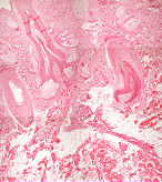
Area
3 is a section through the red part of the lips -
the
 vermillion border. Note that the epithelium
is keratinised but very thin in this region with
capillary loops coming very close to the surface. It
is this superficiality of the blood vessels and
their number which gives the area its red
colouration.
vermillion border. Note that the epithelium
is keratinised but very thin in this region with
capillary loops coming very close to the surface. It
is this superficiality of the blood vessels and
their number which gives the area its red
colouration.
To open the e-Scope, click on one of the demarcated areas in the micrograph below:-