Periodontal ligament/Bone 4
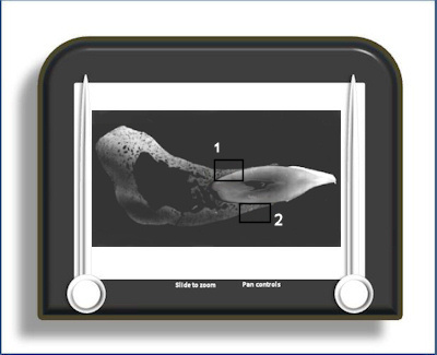
Microradiograph of tooth in situ
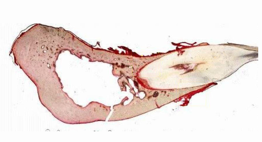
This is a microradiograph (a high resolution x-ray) of a section of a tooth in situ (as seen below)
This a thick, ground, section so there is little if any soft tissue detail. However, the microradiograph provides information about the mineral density of the bone surrounding the tooth - the lighter an area on the radiograph the denser the mineral and vice versa. As can be seen, there is considerable variation in mineral density (and thus maturity of the bone), particularly in area 2. Also visible in area 2 on the periodontal surface of the alveolar bone is a Howship's lacuna where the bone has been partly demineralised. Both areas demonstrate numerous Haversian systems (osteons) with a central canal and concentric layers of osteocytes.
To open the e-Scope, click on one of the demarcated areas in the micrograph below:-