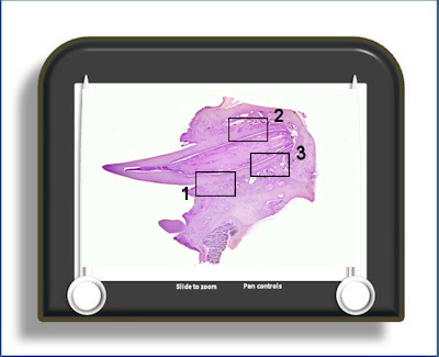
Periodontal ligament/Bone 1
PDL and bone (H&E stain)
This slide is of a tooth in situ with the periodontal ligament showing an area of bone surface where there is bone deposition (area 1), a region of bone resorption (area 2) and a region showing a reversal line (area 3).
The periodontal ligament is the soft connective tissue that attaches the tooth to the bone. It is highly cellular with numerous fibroblasts (occupying up to 50% of the connective tissue volume). The direction of the collagen fibres can be deduced from the orientation of these cells. The ligament also has numerous capillaries which occupy a significant proportion of the periodontal space and which may link to blood spaces in the surrounding alveolar bone via Volkmann's canals
The bone surface in area 1 where there is
bone deposition
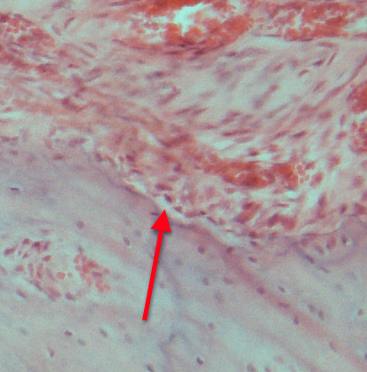 shows a layer of osteoblasts. A
lighter-stained layer of osteoid is evident
between the osteoblasts and the mineralised bone
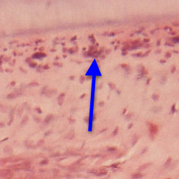
(red arrow). Also evident in area 1 is the
layer of epithelial rests of Malassez
running adjacent to the cementum surface of the
root (blue arrow). The bone surface in area 2
(diametrically opposite area 1) is undergoing resorption in a number of sites. The cells
responsible for this
shows a layer of osteoblasts. A
lighter-stained layer of osteoid is evident
between the osteoblasts and the mineralised bone
(red arrow). Also evident in area 1 is the
layer of epithelial rests of Malassez
running adjacent to the cementum surface of the
root (blue arrow). The bone surface in area 2
(diametrically opposite area 1) is undergoing resorption in a number of sites. The cells
responsible for this
 are osteoclasts which are
large multinucleated cells which often stain
with a more orange/pink hue than surrounding
cells. They sit in (Howship's) resorption
lacunae.
are osteoclasts which are
large multinucleated cells which often stain
with a more orange/pink hue than surrounding
cells. They sit in (Howship's) resorption
lacunae.
Area 3 has a prominent reversal line. This is a 'fossilised' line in the bone that marks a previous resorbing bone surface that has subsequently had new bone deposited on it.
To open the e-Scope, click on one of the demarcated areas in the micrograph below:-