Periodontal ligament/Bone 2
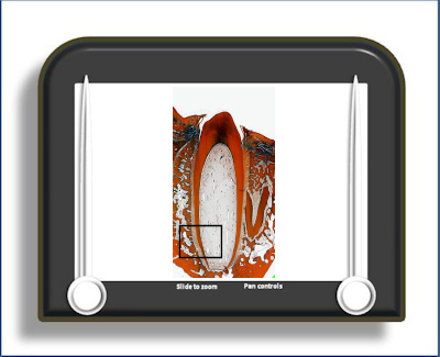
PDL and bone (picric acid stain)
This is a section of a tooth in situ where the periodontal ligament has been stained with picric acid rather than the usual H&E. This preferentially stains the fibre bundles within the tissue and stains the cells poorly, making the orientation and distribution of the fibres more obvious. These fibres within the periodontal ligament enter the bone as Sharpey's fibres and in this section can be traced for some distance within the bone. Bone where the inserting fibres are prominent in this way is termed bundle bone. This appearance is typical of newly deposited bone. As it matures and remodels these fibres become obscured and may eventually be replaced by haversian systems. Note that the Sharpey's fibres where the periodontal fibres insert into cementum are much smaller and more numerous.
In the bulk of the periodontal ligament the fibre bundles seen in relatively thick histological sections like this one appear to be orientated such that they run obliquely down from alveolar bone to cementum.
To open the e-Scope, click on the demarcated area in the micrograph below:-