Tooth Development 4
Root development
This is a section through the
skull of a rat highlighting a mandibular molar
where the root is developing (blue
arrow) (also
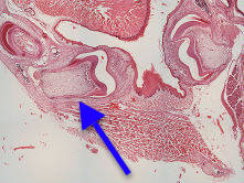 present
are a maxillary molar and an incisor). The crown
shows enamel in various stages of development
from the cusp tip where there is no remaining
enamel matrix to the cervical margin where the
enamel matrix is intact and yet to be degraded.
At the cusp tip the enamel organ is greatly
reduced.
present
are a maxillary molar and an incisor). The crown
shows enamel in various stages of development
from the cusp tip where there is no remaining
enamel matrix to the cervical margin where the
enamel matrix is intact and yet to be degraded.
At the cusp tip the enamel organ is greatly
reduced.
The root sheath is a bilaminar structure derived from the inner and outer enamel epithelia. It has nothing analagous to the stratum intermedium or stellate reticulum (this may be significant in the lack of enamel production on the root). The root sheath induces the outer cells of the papilla adjacent to it to differentiate into odontoblasts and very rapidly to start to produce root dentine. The root sheath is always the same length regardless of how long the root is because once dentine formation is initiated the root sheath disintegrates at its coronal end (resulting in the rests of Malassez). Before disintegration the cells of the root sheath may deposit (enamel) matrix proteins on the dentine surface. This layer may be important in the subsequent induction of cementoblasts in the follicle adjacent to the root.
The periodontal ligament develops from the follicle. In teeth without predecessors (i.e permanent molars) the oblique fibres of the ligament develop at the same time as the root (as in this case of the rat molar). In teeth such as human premolars the oblique fibres do not develop until the tooth has erupted through the gingiva.
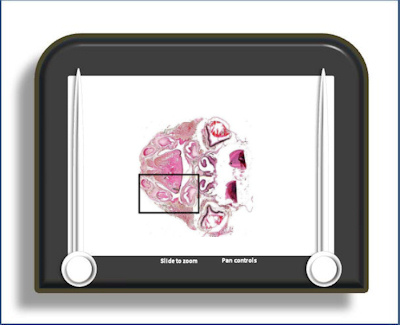
To open the e-Scope, click on the demarcated area in the micrograph below:-