Oral Mucosa 4
Soft palate
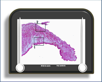
This is a longitudinal section of the soft palate, stained with H&E. The tissue has two 'outer' surfaces covered by an epithelium- a dorsal (nasal) surface and a ventral (oral) surface.
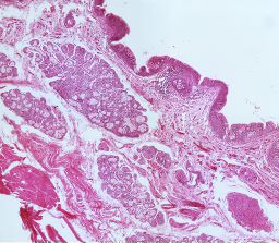
The oral surface
(area 1) is a typical lining mucosa with a
non-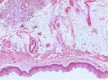 keratinised epithelium
with short, flat epithelial ridges/dermal
papillae. The
lamina propria of the oral mucosa is thinner
than the more anterior parts of the palate (the
hard palate) with an increased number of
capillaries. There is a large sub-mucosa with
prominent minor salivary glands. Deep to the
sub-mucosa is the central core of striated
muscles
keratinised epithelium
with short, flat epithelial ridges/dermal
papillae. The
lamina propria of the oral mucosa is thinner
than the more anterior parts of the palate (the
hard palate) with an increased number of
capillaries. There is a large sub-mucosa with
prominent minor salivary glands. Deep to the
sub-mucosa is the central core of striated
muscles
The dorsal (nasal) surface epithelium (area 2)
is a thin pseudo
 stratified
ciliated columnar epithelium containing goblet
cells and with many minor serous, mucous
and mixed glands beneath it.
stratified
ciliated columnar epithelium containing goblet
cells and with many minor serous, mucous
and mixed glands beneath it.
To open the e-Scope, click on the demarcated area in the micrograph below:-